(Fig.1) Fluorescent microscopes cannot distinguish single atoms.
The 1st, 3rd, 5th paragraphs of this hyped news (7/29/2025) say
"The glycocalyx surrounds each cell in the human body like a coat."
"Nevertheless, it has not been possible to link its spatial organization to its biological function, because it takes place on a scale of only one nanometer, a size that could not be made visible using previous methods." ← The present science intentionally avoids using practical atomic force microscopes manipulating single atoms.
"the researchers combined a special localization microscopy method (Resolution Enhancement by Sequential Imaging, RESI) with bioorthogonal chemistry in which the cell's metabolism is used to attach specific markers to target structures"
↑ This research paper ↓
p.1-abstract says "the visualization of individual sugars within glycans on the cell surface, thus obtaining images of the glycocalyx with a spatial resolution down to 9 Å in an optical microscope" ← This method can Not obtain the atomic level (= 1 Å ) resolution
p.8-left-2nd-paragraph says "Six orthogonal DNA sequences modified with aza-dibenzocyclooctyne (DBCO) at their 5′ ends were used to label the azido sugars"
This-3rd-paragraph says
"Resi involves labelling adjacent copies of the same target molecule with a different tag. The researchers showed that when each of the tags was lit up, they could be clearly differentiated"
↑ This RESI method just labeled some molecule with fluorescent tag ( this-p.23 ) that can be measured by a optical microscope at bad resolution (= 9Å ) that cannot distinguish single atoms (= just 2D vague fluorescent image can be obtained ).
This-p.3-3rd~4th-paragraphs say
"The size of the tags that light up to provide the position information also becomes a limiting
factor at these length scales, as it is the position of the fluorescent part of the tag that is found, not
the interesting part of a molecule it is connected to"
Even the latest research on fluorescent microscope has very bad resolution of 60 nanometers ( this-1st-paragraph, 8/4/2025 ), which is far larger than a single atom of 0.1nm.
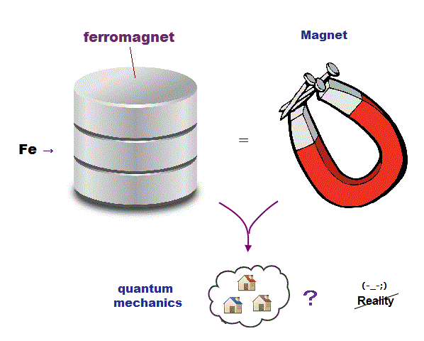
As a result, only practical multi-probe atomic force microscopes can directly observe and clarify atomic structures and functions of cells, membrane proteins and sugars by using artificial chemical bond breaking gradually disassembling the target proteins, which has been hampered by unphysical quantum mechanical model.

Feel free to link to this site.